Showing posts with label Image. Show all posts

Thursday, December 5, 2013

Image post 10: stunning vertebra makes contest list


This image shows a lumbar vertebra from a mouse with its back to the upper-left and its belly to the lower-right. There are muscle fibers in the upper-left corner, cartilage in blue merging into bone in green at the ends of the vertebra, red blood cells in the middle and intestinal contents in orange in the bottom-right corner. The image, which won 13th place in the Nikon Small World Contest, was captured by Dr. Michael Paul Nelson in the Division of Neuropathology, part of the Department of Pathology within the UAB School of Medicine. Dr. Nelson says he created this image, not for a specific research goal, but instead to help him understand aspects of mammalian anatomy. The tissue sample in the image was prepared with a series of dyes and was revealed when Dr. Nelson switched his microscope to fluorescent mode.

When he took the image, Dr. Nelson was attending the Immunohistochemistry and Microscopy Short Course offered by the Histochemical Society at the Marine Biological Laboratory at Woods Hole, MA. Also credited for the Nikon content entry was Samantha Smith, the representative from Carl Zeiss Microscopy who helped Dr. Nelson with technical aspects of using a Zeiss microscope.

Monday, September 23, 2013

Image post 9: infection blocks trash removal to cause ulcers, cancer


What's that ... a meteor blazing past a molten planet?  No, it's a self-destructing cell just shed from a gastric gland made up of the tightly packed blue cells running across the bottom.

Gastric glands that secrete digestive juices into the stomach, like all epithelial cells (skin, lining of blood vessels, etc.), constantly shed old cells from their outer layers and replace them with new ones. The turnover keeps tissues viable throughout adult life. When the shed cells sense they have outlived their usefulness, they initiate self-destruct mechanisms.

With some cells always in the process of self-destructing, other nearby cells are charged with swallowing up the dying cells and disposing of them. That explains the red cloud surrounding the yellow dot at the center of the image. An antigen-presenting cell (dyed red) has engulfed a self-destructing cell (yellow) to remove it.

The system works pretty well until a person's stomach gets infected with the bacteria Helicobacter pylori.

The infection causes cells to release TNF-alpha, a signaling chemical that triggers the waves of cell activation and chemical release meant to kill invading bacteria or viruses (inflammation). While the process protects us against infectious disease, it also plays a role in many disease processes when it goes too far.

recent study by a team of UAB researchers found that, along with triggering inflammation, TNF-alpha also blocks the engulfment and removal of dying cells. Cellular debris builds up to drive further inflammation in a vicious cycle.

Why does this matter?  Inflammation caused by Helicobactor pylori is behind the development of nearly all ulcers. Furthermore, the same chronic inflammation damages DNA, creating risk for hard-to-treat gastric cancer.

The image was created by Diane Bimczok, DVM, Ph.D., an instructor in UAB's Division of Gastroenterology and Hepatology, using routine fluorescence microscopy and digital imaging. Phillip Smith, M.D., was senior author of the related study. 

Wednesday, August 21, 2013

Image post 8: mapping live brains

This image from UAB Research illustrates the improving brain maps that promise to reveal the mechanisms behind complex, neurological diseases.


A MRI technology called diffusion tractography captured this image of a living rat brain. Tractography is a 3-D modeling technique that visually represents nerve pathways in the brain using data collected by diffusion tensor imaging (DTI).

DTI visualizes nerve pathways known collectively as white matter that connect the various parts of the brain via long bundles of nerve cells. It shows whether or not bundles of white matter fibers run in the same direction, but not in which direction. Tractography does that.

Since developed by researchers at Washington University School of Medicine in St. Louis, it has given the field a more detailed look at brain structures, especially living brains. Taken together, these new MRI technologies promise to improve understanding of neurological disorders like schizophrenia and Alzheimer's disease and the consequences of head trauma. They also may one day help neurosurgeons better avoid cutting nerves during surgeries.

In particular, tractography reveals connections that can be measured in living human subjects and measurements that can be made simultaneously across the entire brain. The Human Connectome Project is capitalizing on these strengths to build more accurate brain maps. The image was created in the lab of Hyunki Kim, Ph.D., associate professor in the departments of Radiology and Biomedical Engineering and faculty in the UAB Comprehensive Cancer Center.

Friday, July 26, 2013

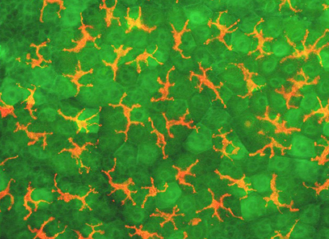
Image post 7: spectacular skin cells

Another interesting image coming from UAB research captures the skin's ability to turn back the sea of microbes surrounding it.   


Pictured here is an outer cell layer of mouse skin. Humans have something similar. The tough, fibrous protein keratin, which gives structure to skin, has been dyed green, and T cells that watch for viruses, bacteria or parasites seeking to invade the body have been dyed orange.

These particular T cells, called gamma delta T cells, are unique in their spectacular appearance. They have been described as a missing link between the more primitive innate immune system, which is quickly, directly activated by foreign invaders, and the slower but more precise adaptive system, which must first be first primed by precise mechanisms before it can unleash clonal cell armies specific to the invader at hand.

Gamma delta T cells do a bit of both, and researchers seek to better understand how they defend the skin from invading microbes and help heal wounds. The image was generated in the lab of John Kearney, Ph.D., professor in the Department of Microbiology within the UAB School of Medicine.

Wednesday, June 26, 2013

Image post 6: spinal cord cell reaches for its neighbor

While many posts from The Mix feature a science story, we also share images coming out of UAB research. Below is a description of what we are looking at and related hints about how the brain forms in the womb. 


Pictured here is one star-shaped astrocyte "reaching out" to another in a dish. The most abundant cell type in the brain and spinal cord, astrocytes are not nerve cells, but instead provide support, nutrients and protection to nerve cells. Recent work has shown that astrocytes help to shape the messages being passed from nerve cell to nerve cell, and that problems with astrocyte function may throw off nerve cell performance.

In her research, Michelle Olsen, Ph.D., assistant professor in the UAB Department of Cell, Developmental and Integrative Biology, seeks to determine how the overlap between nerve cells and astrocytes contributes to normal brain development, and to brain abnormalities when something goes wrong.

Dr. Olsen's experiments with isolated cells seek to model processes underway in the brain as it forms during development. Nerve cells are known to put out "roots" that reach out, find nearby cells and link up to form signaling networks. The above picture suggests that astrocytes do something similar.

Named for their star shape, astrocytes put out extensions that wrap around synapses, the gaps between nerve cells in signaling pathways. Each nerve cell in a pathway sends an electric pulse down itself until it reaches a synapse, a gap between itself and the next cell in line. When it reaches the cell's end, the pulse triggers the release of chemicals called neurotransmitters that float across the gap. Arriving at the other side, they cause the downstream nerve cell to “fire” and, depending on the synapse type, to either pass on or stop the message.

In this way, each synapse between nerve cells “decides” whether or not a message continues down that pathway. The balance of messages passed on (excitation) and messages halted (inhibition) is crucial to brain function. One theory has it that astrocytes influence that balance at synapses with their own set of extensions and transmitters.

Dr. Olson captured the image using an inverted Zeiss Observer microscope. 

Thursday, May 30, 2013

Image post 5: eye nerves shed light on memory disorders

While most posts from The Mix feature a science story, we have also begun sharing images coming out of UAB research. Below is a brief description of what we are looking at and how related work may contribute to a better understanding of Alzheimer's disease.


Pictured here is a retinal ganglion (center), a kind of nerve cell near the eye’s retina that helps to process light into the images we perceive. It had been injected with a fluorescent dye, which made it glow green along with the cells connected to it electrically. In each of our eyes, 125 million photoreceptors capture light. They then trigger nerve messages in 1.5 million retinal ganglion cells, long extensions of which bundle together to form the optic nerve.

Captured by Christianne Strang, Ph.D., research instructor in the Department of Vision Sciences within the UAB School of Optometry, this image represents signaling mechanisms between the retina and surrounding nerve cells. Strang's lab seeks to understand how photoreceptors connect to surrounding nerve pathways, as well as the degree to which they signal using the neurotransmitter acetylcholine.

Within nerve pathways, each nerve cell sends an electric pulse down an extension of itself called an axon until it reaches a synapse, a gap between itself and the next cell in line. When it reaches an axon’s end, the pulse triggers the release of chemicals called neurotransmitters that float across the gap. Upon reaching the other side, they either cause the downstream nerve cell to “fire” and pass on the message, or stop the message. Certain neurological diseases, including Alzheimer’s, have been linked to a decrease in acetylcholine signals in nerve pathways related to vision and memory.

As for rest of the color scheme, the pictured eye tissue has also been treated with dyes that interact with choline acetyltransferase (blue), which helps to produce acetylcholine, and synaptophysin (red), which reveals the location of synapses. The work was done in the lab of Kent Keyser, Ph.D., professor in the School of Optometry.

Thursday, May 16, 2013

Image post 4: nanodiamonds may solve implant problem

While most posts from The Mix feature a science story, we have also begun sharing images coming out of UAB research. Here is an image of nanodiamonds currently being studied as a potential coating for artificial joints. UAB researchers are exploring whether such coatings can reduce wear on joints made of metal alloys. The work is important because more than 400,000 knee replacements and 300,000 hip replacements are performed each year in the United States.

The grinding force placed on joints causes the artificial versions to shed debris that can cause pain, limit mobility and hasten joint failure. Debris particles are absorbed by scavenging immune cells called macrophages, which then secrete chemicals that cause swelling and pain. This inflammation turns on bone-eating cells near implants, and bone-loss increases the likelihood implants will break loose and require a second surgery.

Diamond coatings may significantly reduce such shedding, and studies are underway to confirm that they are safe and effective. For more information on the work led by Yogesh Vohra, Ph.D.,  director of the UAB Center for Nanoscale Materials and Biointegration, read this 2012 article.


Wednesday, May 8, 2013

Image post 3: dangerous clumps of fungus

While most posts from The Mix feature a science story, we have also begun sharing images coming out of UAB research. Below is a brief description of what we are looking at and how related work may help to diagnose and treat fungal infections.


Here is a scanning electron microscope image of the fungus called Aspergillus. It's in the process of germinating, or emerging from round spores (at the center) to begin growing. The fungus has sprouted long, branching filaments called hyphae.

Most people breathe in Aspergillus spores daily without incident, but those with lung diseases or weakened immune systems can contract Aspergillosis, symptoms of which range from allergic reactions to severe lung infections. The fungus is a major player in some forms of allergic asthma, as clumps of hard-to-remove hyphae build up in the lungs.

According to the CDC, fungal infections pose an increasing threat to public health because of the growing number of people with weakened immune systems, including AIDS, cancer and transplant patients. In addition, treatment-resistant fungal infections have emerged as a growing problem in hospitals. Global warming may be contributing to an increase in infections, as fungi thrive in warm, moist conditions. Please see the CDC fungal page for more.

Current treatments are largely incapable of reducing morbidity and mortality in Aspergillosis, said John Kearney, Ph.D., professor in the Department of Microbiology within the UAB School of Medicine. He and his team are developing a new kind of vaccine that could provide protection against invasive Aspergillosis. Bacteria elicit a stronger human immune response than fungi but contain some of the same proteins (e.g. chitin). Based on these common building blocks, it may be possible to develop a vaccine where bacterial protein vaccine ingredients are used to activate immune cells that also target a fungus and remove it from the body.

This image was made by Dr. Jeffrey Sides from the Kearney laboratory at UAB using an instrument made available by the UAB School of Engineering.


Wednesday, May 1, 2013

Image post 2: Worm gonads and Lou Gehrig's disease

While most posts from The Mix feature a science story, we have also begun regularly sharing images coming out of UAB research. Below is a brief description of what we are looking at and how related work may help diagnose and treat Lou Gehrig's disease.  As we get more of these, we will add them to a slide show on the blog page and share them via FacebookTumblr and Pinterest.


This image is a dissection of a species of worm called  Caenorhabditis elegans (C. elegans), among the most famous of worms because it has made possible several discoveries in molecular biology. It did so by serving as a simple model of cellular processes conserved by evolution and still at work in humans.

The picture shows the worm's intestine running across the middele (in blue), which is working to provide fatty building blocks (e.g. omega-3 polyunsaturated fatty acid) to the worm's egg-producing cells (oocytes) in green. The egg-producing cells then convert the fatty acids into chemical cues called prostaglandins that help sperm find the sites (purple areas) where they can fertilize the eggs. C. elegans sperm in turn release a protein called major sperm protein (MSP), which tells the egg to prepare for fertilization.

Interestingly, the studies related to this image are providing key insights into how prostaglandins and MSP are made and function in humans.While MSP, for instance, was first found in worms and in connection with reproduction, it appears to have been put to work by human evolution in signaling roles in many cell types.

For instance, Michael Miller, Ph.D., associate professor in the UAB Department of Cell, Developmental and Integrative Biology, and creator of the attached image, last year published key work showing that MSP may be involved in the development of amyotrophic lateral sclerosis, or Lou Gehrig's disease. This unexpected connection may provide new approaches to diagnosing and treating the disease.

Note: if you have an amazing UAB research image you would like to share, please email mwindsor@uab.edu.

Wednesday, April 24, 2013

Image post 1: brain message superhighways

While most posts from The Mix feature a science story, we also wanted a forum to share powerful images coming out of UAB research. Regular image posts will be accompanied by a brief description, including how the image depicted might soon be important to science or medicine. The post will also link to the creator of the image.


Above is a face view of a brain’s white matter created by UAB graduate student Meredith Reid using an MRI technology called diffusion tensor imaging (DTI). The strands running through the image are axons, long extensions of nerve cells that form pathways carrying messages between the parts of the brain. The colors represent the spatial orientation of the axons, with one color for those running left to right, another for those running back to front, etc. Certain qualities of such images give researchers a measure of the integrity of white matter axon fibers, which promises to improve understanding of neurological disorders like schizophrenia. The work was done in the lab of Adrienne Lahti, M.D., professor in the Department of Psychiatry and Behavioral Neurobiology within the UAB School of Medicine. On a final, related note, it's also worth checking out the Human Connectome Project run by the National Institutes of Health.  

Note: if you have an amazing UAB research image you would like to share, please email mwindsor@uab.edu.